
Table of Contents
Introduction — a quick scene, some numbers, a sharp question
Have you watched a blood flow map and felt a little cheated? I have. The room is quiet. The monitor glows. The data says change, but the patient looks the same. laser speckle contrast imaging lsci shows flow in real time — but does it tell the whole truth? (Small labs, big hopes.)
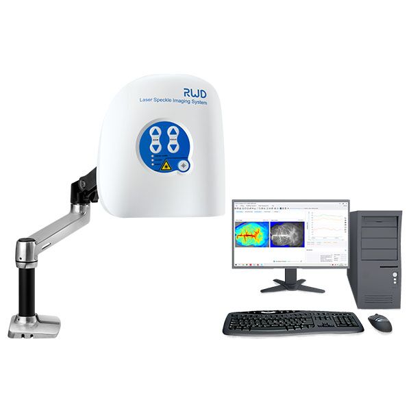
Here I set a simple scene: a neurosurgeon needs perfusion feedback during a case. The camera gives 30 frames per second, and the lab logs a 20% drop in perfusion index. Yet the surgeon hesitates. Why the pause? Why trust or doubt the image? That doubt is the central question we will explore next — and it matters for practice, for devices, for patients.
Peeling Back the Layers: Where Traditional Methods Fail
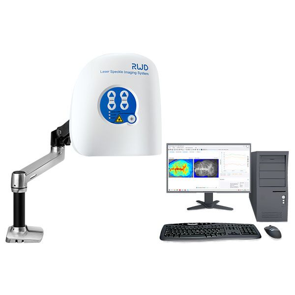
laser speckle contrast imaging promises quick maps of microvascular flow. I’ve used it in the OR. I know its strengths. I also know the common failures. First, raw speckle contrast depends on camera sensor specs and frame rate. Second, signal-to-noise ratio can wobble with motion. Third, simple algorithms assume steady illumination. These are not minor issues — they shape conclusions. Look, it’s simpler than you think when you break it down.

Why does it break?
Most systems use fixed exposure and a single processing path. When the patient moves, or when ambient light shifts, the speckle contrast shifts too. The result: false hotspots or muted flow. I’ve watched a perfusion map swing while the surgeon adjusted a light — funny how that works, right? Edge computing nodes or local GPUs can help, but many setups still rely on raw camera output and naive averaging.
There’s also the human side. Clinicians expect clear thresholds. But speckle contrast is relative. The math plays with decorrelation time and exposure. Without calibration, you get numbers that feel authoritative but are context-dependent. My point: the traditional solution is brittle. It treats complex signals as if they were simple. That mismatch creates real-world trouble for diagnosis and for device validation.
Looking Forward: Principles and Practical Steps
Now I shift a bit. Let us think about what comes next. I favor approaches that combine better optics, smarter processing, and clearer user feedback. New systems must adapt exposure on the fly. They should use multi-exposure stacks to separate motion from flow. When I test prototypes, I watch for stability over clamps of different durations and for consistency across camera models.
What’s Next?
One path is hybrid processing: blend real-time speckle analysis with short-term averages and motion compensation. Another is to add simple calibration curves so a clinician sees confidence bounds, not just colors. I’ve seen prototype instruments apply on-device filtering and deliver cleaner maps in under a second — results that the team trusts more. The future is iterative — small changes that reduce false positives and improve clinical adoption.
From my view, three practical metrics matter when you evaluate a solution: temporal fidelity (can it track rapid changes?), robustness to motion (does it resist artifacts?), and interpretability (are outputs easy for clinicians to use?). Use those to compare systems. I also recommend field trials; no lab test replaces a 10-case OR run. We need transparency in processing steps. If a vendor hides post-processing, I ask questions. I want traceability — and so should you.
To close, I’ll be frank: technology helps, but so does humility. We must pair tools with clear training and honest reports of limits. There is promise in adaptive speckle methods — and real work ahead. For more on practical systems, I look at the options from companies like BPLabLine, and I test for real-world fit before I trust a map with a patient’s fate.
